Cone beam CT is not a routine film. Here are the situations where it genuinely changes the plan, and how it differs from a normal x-ray.
Cone beam computed tomography, usually shortened to CBCT, produces a three dimensional image of the jaws. Structures that overlap on a normal radiograph are separated here: bone width can be measured in millimetres and the path of a nerve canal can be followed. It is not a routine film. It is requested when a two dimensional image cannot answer a specific question.
The usual indications
- Implant planning, where bone width, height and nerve position must be measured
- An impacted wisdom tooth whose root appears close to the nerve canal
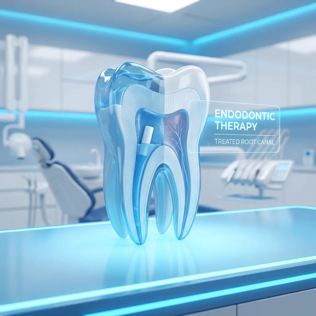
- A root canal that failed, with a suspected extra canal or a vertical fracture
- A lesion in the jaw with unclear borders
- Surgery planned close to the floor of the sinus
- Trauma where a fracture line is not clear on a plain film
Why implants rely on it
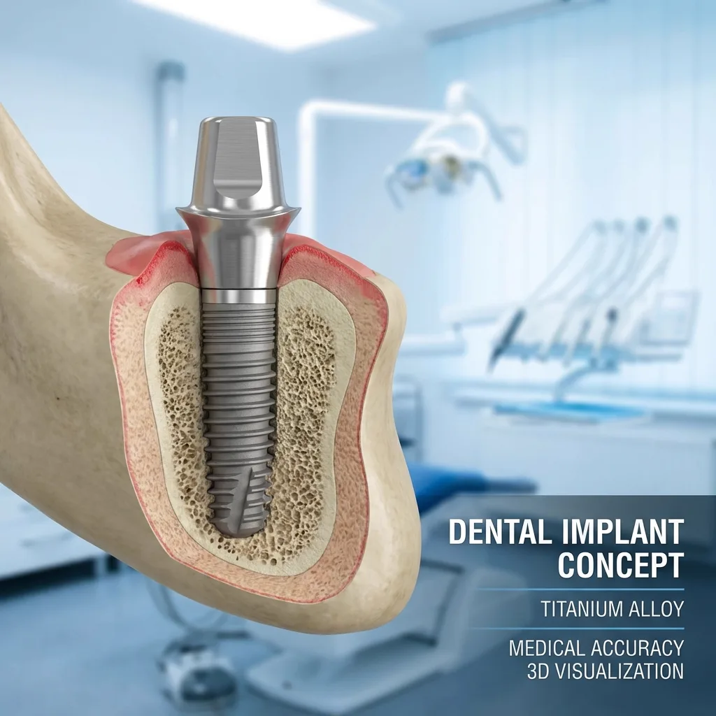
A panoramic film shows bone height but not bone width, because everything is projected onto one plane. Cross sections show the site from the front and from the side, and those measurements decide the diameter, length and angle used in implant treatment, or whether bone grafting is needed first.
Safety before surgery
When a root and a nerve canal overlap on a flat image, that does not prove they touch. It only proves they line up. A cross section shows whether the root passes in front of the canal or behind it, and in cases that need oral surgery that single fact can change the whole approach.
How the scan is taken
You rest your chin on a support, the machine makes one rotation around your head and the scan is usually over in less than half a minute. Nothing is placed inside your mouth. Staying still matters, because even slight movement blurs the slices.
Dose and justification
A CBCT carries a higher dose than a single small film, so it should always have a reason behind it and the scanned field should be kept as small as the question allows. You can ask your dentist what the scan will change in the plan. For a routine check up or a simple filling, it is not needed.
Practical points
- Metal fillings and crowns create bright streaks that can obscure neighbouring areas
- Jewellery, glasses, hairpins and removable dentures come off before the scan
- A scan taken too soon after an extraction shows immature bone and can mislead
- An old scan is still worth bringing, since it shows how the bone has changed
- Reading the slices is a skill in itself; an image on its own is not a diagnosis