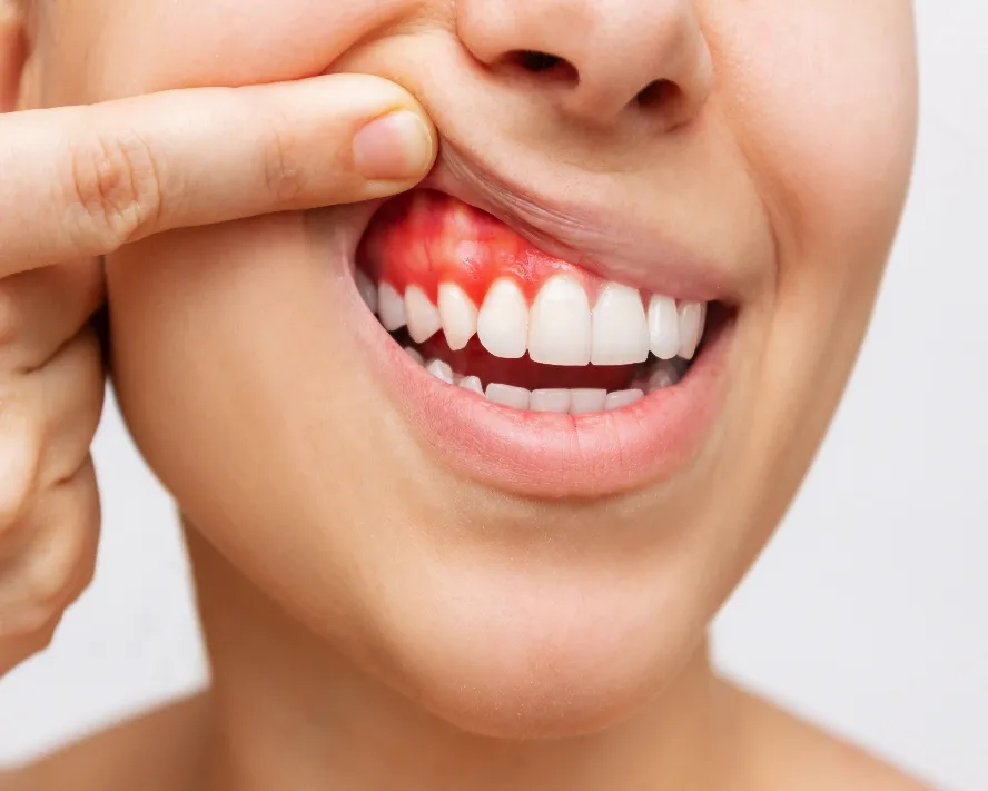
Tongue and lip piercings can cause chipped teeth and gum recession over time. The risks, the early warning signs, and how to protect the mouth if you keep one.
Oral piercings are seen as an aesthetic choice, and most people have one placed without asking a dentist. A metal object sitting in the mouth for years, though, leaves an effect on teeth and gums that accumulates quietly. Those effects are mostly irreversible.
Chipped teeth are the commonest problem
The metal ball of a tongue piercing knocks against the teeth continually during speech and swallowing. Each contact looks harmless on its own, yet it repeats hundreds of times a day. Micro-cracks form in the enamel first, then visible chips.
The teeth most affected are the lower back molars, where the tongue naturally rests, and the back surfaces of the upper front teeth. The risk is higher still in teeth with crowns or fillings; porcelain is more brittle than natural enamel.
Gum recession and bone loss
Lip and lower-lip piercings press continually against the gum with their back plate. Gum under constant mechanical pressure recedes, and the bone beneath follows that recession.
The area most often affected is the front of the lower incisors. The gum there is already thin and the bone beneath it narrow; there is no reserve to compensate for loss. Receded gum does not return on its own.
Early warning signs
- Sensitivity in the tooth the piercing touches
- Small notches or roughness at the edge of a tooth
- Redness and bleeding in the gum around the piercing
- The lower front teeth appearing longer
- Persistent swelling or discharge at the site
Once these appear, damage has begun. Early intervention can stop progression but does not recover what has been lost.
The risk of infection
The mouth is a bacterially rich environment, and a fresh piercing is an open wound. Swelling in the first weeks is an expected response; with a tongue piercing it can rarely reach a level that compromises breathing.
Even after healing, the area around the piercing remains a favourable site for plaque accumulation. In patients with heart valve disease or a suppressed immune system this risk needs separate consideration.
Metal allergy and discolouration
Nickel-containing alloys can cause redness, itching and persistent inflammation at the contact site in sensitive individuals. Titanium and surgical steel are safer options but not entirely without risk.
Prolonged contact with metal can leave a grey-blue discolouration in the gum. It remains after the piercing is removed and requires a separate procedure to address.
If you are keeping it
For patients who do not want to remove the piercing, we suggest a practical protection plan. The most effective step is replacing the metal ball with an acrylic or plastic one; that single change markedly reduces the risk of chipping.
- Use the shortest barbell length that fits
- Do not play with it against the teeth while speaking or idle
- Check screw ends regularly; they can be swallowed
- Clean around the piercing at every brushing
- Remove it during sport
- Attend a check-up every three months rather than every six
What happens when it is removed
The hole usually closes within weeks, often leaving a small mark on the tongue. Gum recession and chipped teeth, however, do not correct themselves. Removing the piercing stops the damage advancing; it does not undo it.
The earlier the decision is made, the less intervention the situation needs. With a piercing worn for years, receded gum may need grafting and chipped teeth composite repair.
When to have it looked at
The most sensible route for someone considering a piercing is an examination beforehand; the thickness of the gum and the arrangement of the teeth can be assessed for how they affect the risk.
With younger patients, having this conversation as information rather than prohibition changes the outcome. A patient who decides knowing the risks is far more willing to follow protective measures; one who is simply refused tends not to return at all.
For those who already have one, regular review is enough. Gum levels are measured and photographed at the examination, so that any progression is followed by measurement rather than guesswork.