




































What Is a Dental Implant and How Does It Work?
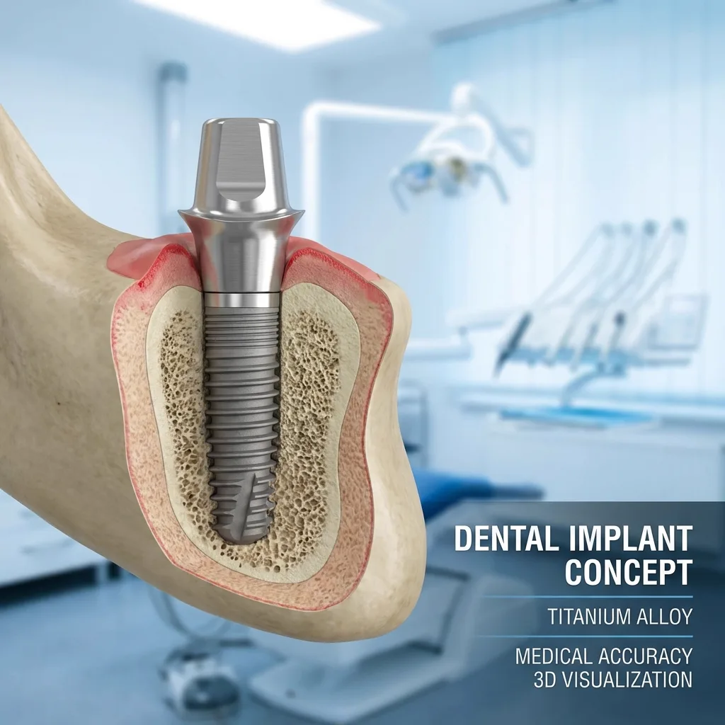
An implant has three parts, each with its own function — which also explains why treatment proceeds in stages.
The three parts of an implant
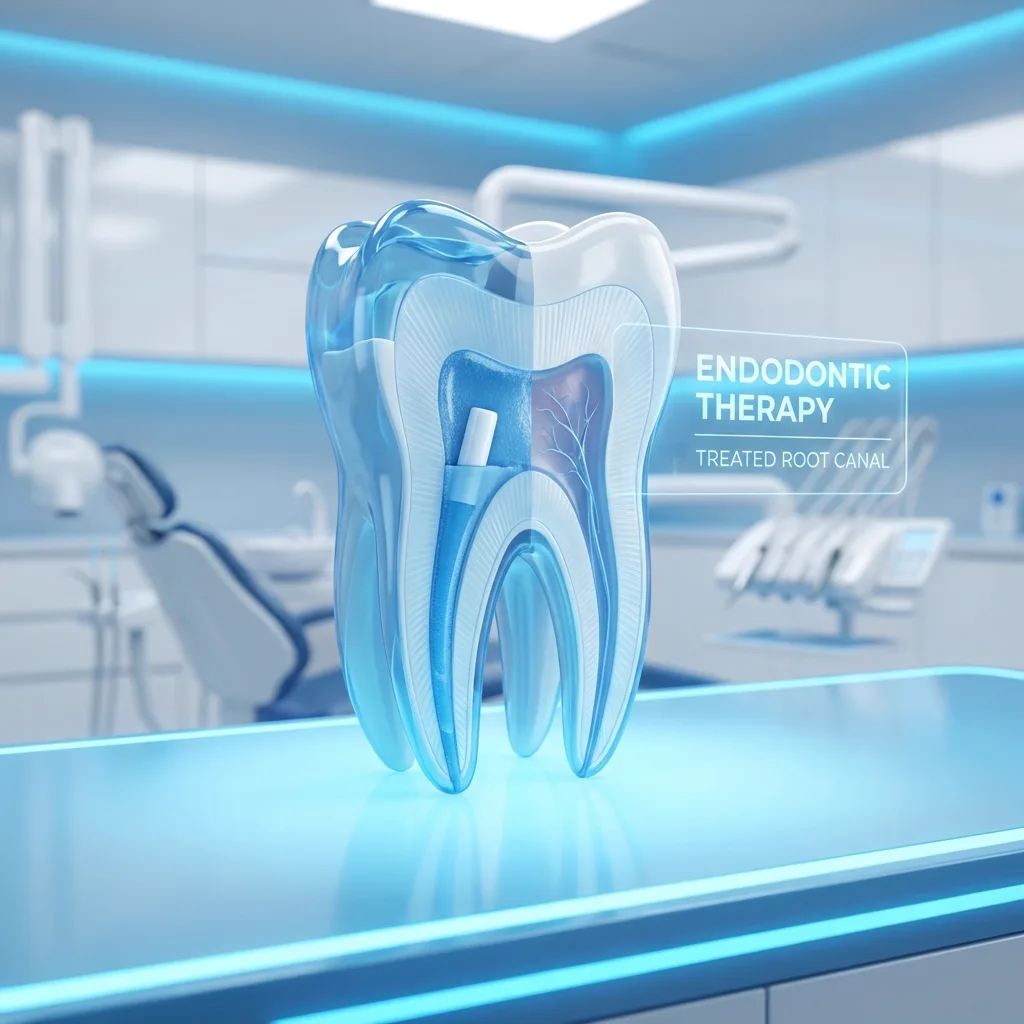
The implant body is the titanium screw in the bone, mimicking the natural root. The abutment connects it to the restoration. The crown is the visible tooth. Because they are separate, only the crown needs replacing if it wears.
Osseointegration: fusing with the bone
Bone does not recognise titanium as a foreign body and grows directly onto it — a biological fusion called osseointegration, usually taking two to four months. Loading it before this completes is the most common cause of failure.
Implant, bridge or denture?
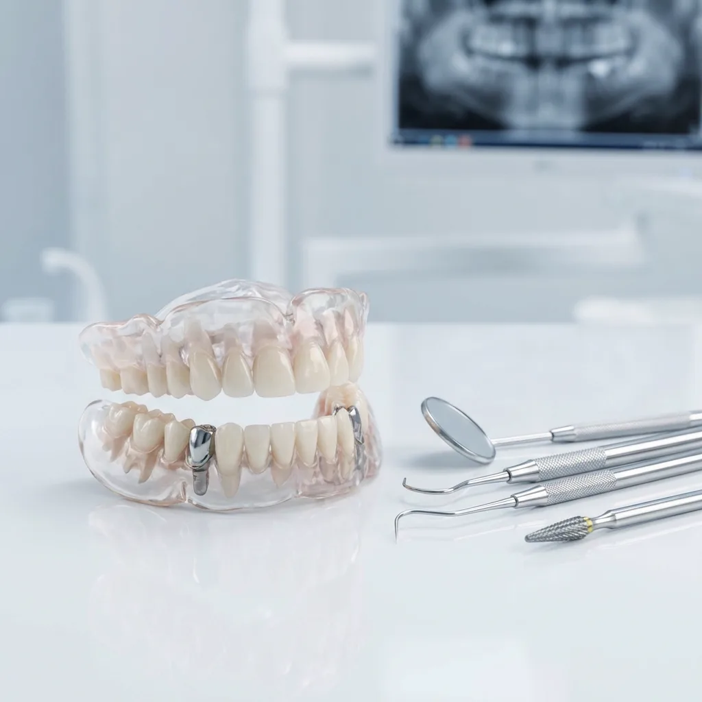
For a single gap the implant is clearly superior. For multiple gaps an implant-supported bridge is far more comfortable than a removable denture, with no palatal coverage. In complete edentulism, implant support eliminates slipping.
How Implant Treatment Proceeds in Antalya
Treatment is a planned process and no stage is skipped; total time depends on bone condition and whether additional procedures are needed.
1. Examination and digital planning
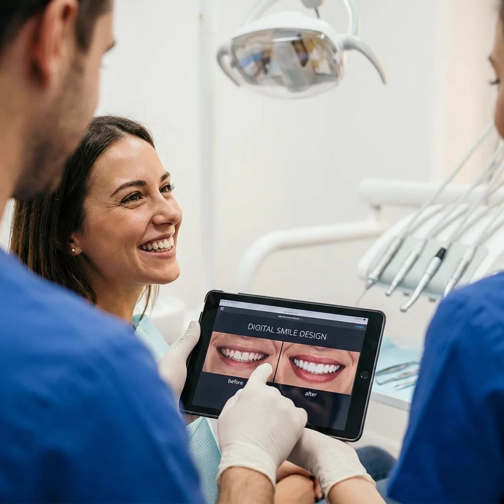
An intraoral examination and panoramic X-ray assess bone height, width and density, and the distance to the nerve canal or sinus. These measurements determine the implant's diameter, length and angle.
2. Surgical placement
Performed under local anaesthesia, usually 30–60 minutes for a single implant. You feel pressure and vibration but not pain. Some swelling for a few days afterwards is normal.
3. Healing and integration
Two to four months are needed for integration, with a temporary restoration if aesthetics require it. Avoiding smoking is critical, as it impairs microcirculation and is the most common preventable cause of implant loss.
4. The restorative stage
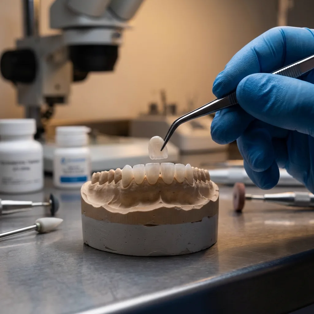
Once integrated, a healing abutment shapes the gum, then an impression is taken and a custom crown made — zirconia or porcelain depending on the site and aesthetic requirements.
Who Is Suitable for Implants?
Implants are suitable for most adults, though some conditions require preparation first or point to alternatives.
Suitable candidates
Good candidates have completed jaw growth, sufficient bone volume, healthy gums and consistent oral hygiene. There is no upper age limit; the lower limit is completed jaw growth.
Conditions requiring preparation first
Insufficient bone may need grafting or sinus lifting, extending the timeline but making implants possible. Active gum disease must be treated first, and grinding managed with a night guard.
Conditions that increase risk
Uncontrolled diabetes delays healing; heavy smoking markedly reduces success. Bisphosphonates, radiotherapy to the jaw and some immune conditions need separate assessment — always disclose these.
Aftercare and Longevity of Implants
The lifespan of an implant depends as much on aftercare as on the treatment itself.
Peri-implantitis: the main threat
Implants do not decay, but the surrounding tissues can become diseased. Peri-implantitis is plaque-related inflammation that leads to bone loss and eventually implant loss — the implant equivalent of periodontitis.
Daily care
Brush twice daily and clean interdentally with floss or interdental brushes. A water flosser is effective around implants. Avoid stiff brushes and abrasive pastes.
Regular check-ups
Six-monthly reviews measure pocket depth, monitor bone level radiographically and provide professional cleaning. Early peri-implantitis can usually be halted; lost bone does not return.
Assessment and Treatment Planning Before Implant Surgery
Where an implant will be placed, at what angle and in what dimension is decided during planning, not on the day of surgery.
Why Radiographic Assessment Comes First
An intraoral examination shows the width of the edentulous area and the thickness of the gum, but the height and thickness of the bone and the position of the anatomical structures inside it can only be understood through imaging. A panoramic film gives a general map, while three-dimensional tomography provides a cross-section of the bone and makes it possible to select the length and diameter of the implant.
The same imaging also reveals the position of the nerve canal in the lower jaw and the sinus cavity in the upper jaw. When planning leaves a safe distance from these structures, surgery becomes predictable. It also allows an early conversation about whether an additional procedure such as grafting will be needed where bone volume is insufficient.
The Role of Medical History and Medication Use
Because an implant heals inside bone, the patient's general health is as decisive as the surgical technique itself. Blood sugar control, blood thinners, medication used against bone loss, previous head and neck radiotherapy and other regular medication all directly affect planning.
When this information is shared in full, treatment proceeds safely. Where necessary your dentist contacts the physician following you; if a change in medication is required, that decision is made together with the relevant specialist rather than by the dentist alone. A systemic condition is usually not an obstacle to treatment, it simply calls for more detailed planning.
What Should Be Completed Before Surgery
- Treating gum inflammation and establishing an oral hygiene routine
- Restoring decay and broken fillings on the remaining teeth
- Extracting non-restorable teeth and allowing the site to heal
- Measuring bone height and width with three-dimensional imaging
- Evaluating the bite and any clenching or grinding habit
- Recording all medication and systemic conditions
- Deciding on the type of prosthesis to be built on top from the outset
Choosing Between an Implant, a Bridge and a Removable Prosthesis
There is no single way to replace a missing tooth; the choice is shaped by the condition of the neighbouring teeth, the bone volume and the person's hygiene habits.
When a Fixed Bridge Comes Into the Picture
A bridge is made by reducing the teeth on either side of the gap and placing a joined restoration over them. If those neighbouring teeth already have large fillings, fractures or root canal treatment, crowning them creates no additional loss and a bridge can be a reasonable solution.
If, however, the neighbouring teeth are intact and untouched, healthy tooth tissue must be cut to build a bridge. This is where an implant stands out: it completes only the missing area without involving the neighbouring teeth and keeps stimulating the bone. The decision is made by looking at which option achieves the result with less loss of tissue.
Where a Removable Prosthesis Fits In
A removable prosthesis remains a valid option where many teeth are missing and bone support is limited. Being removable makes cleaning easier and it offers a solution for large gaps in a short time.
On the other hand, a removable prosthesis rests on the gums and the remaining teeth rather than on bone; as the bone resorbs over time its fit changes and periodic relining or renewal may be needed. For many people the most suitable path is a combination of the two: designs supported by a small number of implants stay in place yet can still be removed for cleaning.
Topics to Discuss When Making the Decision
- Whether the neighbouring teeth are intact or already restored
- The height and width of bone in the missing area
- The state of gum health and daily cleaning habits
- How many stages the treatment takes and how long it lasts
- Smoking and systemic conditions that affect healing
- Whether the solution can be extended if further teeth are lost
- The ability to attend follow-up appointments regularly