Most white patches in the mouth are harmless, but some need monitoring. Why the difference between a patch that wipes away and one that does not matters so much.
A patient who sees a white area in the mirror usually swings to one of two extremes: ignoring it entirely or assuming the worst. The truth lies between; the great majority of white patches are harmless, but they are not all the same and some need monitoring.
The first distinction: does it wipe away?
This single question does half of the assessment. A whiteness that lifts when wiped gently with gauze or a finger, leaving a red, easily bleeding surface beneath, is usually a fungal infection.
Whiteness that does not wipe away and is adherent to the tissue belongs to a different group, and its diagnosis requires examination. Lesions in this group need monitoring; waiting for them to disappear on their own is not the right approach.
Wipes away: oral candidiasis
Candida is normally present in the mouth and multiplies when immunity or the balance is disturbed. It is common in denture wearers, in dry mouth, in asthma patients using inhaled steroids, in diabetes, and after a course of antibiotics.
Treatment is with antifungal medication, but the real issue is finding the cause. Without correcting denture hygiene, rinsing the mouth after an inhaler or controlling blood sugar, it returns.
Patches that do not wipe away
Several different conditions sit under this heading and need to be told apart. Some are entirely harmless; others are lesions requiring regular monitoring.
- Frictional line: a thin white line inside the cheek along the bite plane
- Morsicatio: an irregular white area from a cheek or lip biting habit
- Lichen planus: lacy white striations on both cheeks
- Leukoplakia: a white plaque that does not wipe off and has no other explanation
- Nicotine stomatitis: a whitened palate with red dots
Why leukoplakia matters
Leukoplakia is a description rather than a diagnosis: a white plaque that does not wipe off and is not explained by another condition. Its importance lies in the fact that a small proportion can show cellular change over time.
When leukoplakia is found the lesion is measured, photographed and reviewed at regular intervals. A biopsy is taken where the surface becomes rough or thickened, contains red areas or the lesion enlarges. Follow-up is the part that must not be neglected.
Risk factors
Smoking and tobacco use in any form is the strongest factor, and the risk multiplies when combined with alcohol. Tobacco products held in the mouth produce marked lesions where they sit.
Chronic mechanical irritation should not be overlooked either: a sharp tooth edge, a broken filling or a denture that continually rubs can produce lasting change in the same area over years. Because these causes are correctable, they are addressed first.
When to attend without delay
Certain findings alongside a white patch bring the assessment forward. Lesions mixing red and white areas, a firm base, bleeding, numbness and a non-healing sore lasting more than two weeks are foremost among them.
A firm lump in the neck, a sense of catching on swallowing and a change in the voice are also findings not to postpone. Their presence does not mean bad news; it means prompt assessment.
What happens at the examination
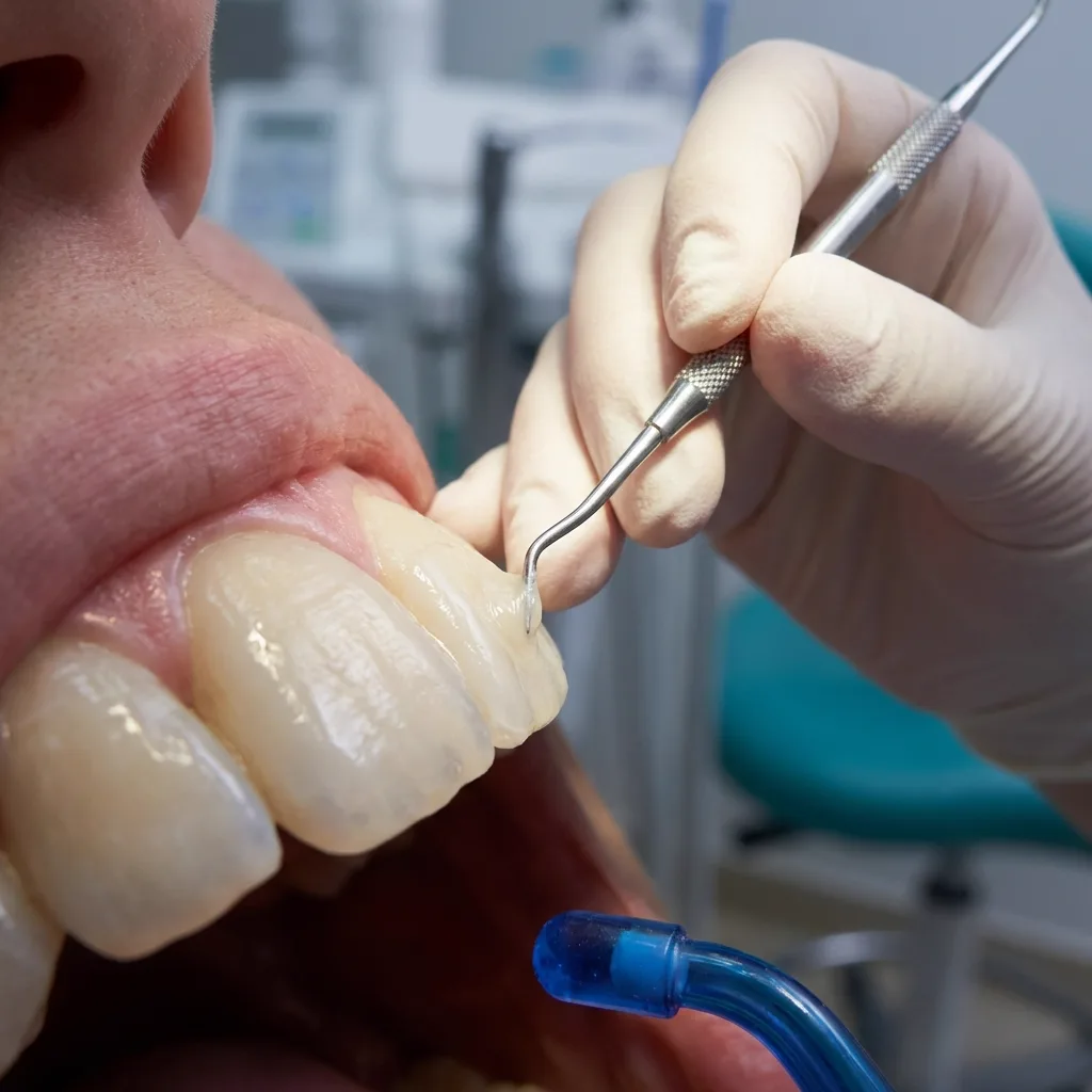
The mouth is examined systematically: the dorsum and borders of the tongue, the floor of the mouth, the cheeks, the palate and the lips. The margins, surface character and firmness of the lesion are recorded, and whether it wipes off is tested.
Where there is an obvious cause — a sharp tooth, an ill-fitting denture — that is corrected first and the area reviewed two weeks later. If the lesion has not regressed, further assessment and biopsy where needed are arranged. This two-week observation is a reasonable route that avoids both unnecessary procedures and delay.
Checking yourself
Looking inside the mouth once a month in good light is a practical habit. Put out the tongue and check its borders, underneath it, the inner cheeks and the inner lips in turn. The aim is not to diagnose but to notice change early.
Anything newly appeared, enlarging or changing colour can be photographed and shown at a review appointment. This screening is carried out anyway at six-monthly examinations — one of the quiet benefits of attending regularly.