











































Gum Aesthetics — The Less Discussed Half of a Smile
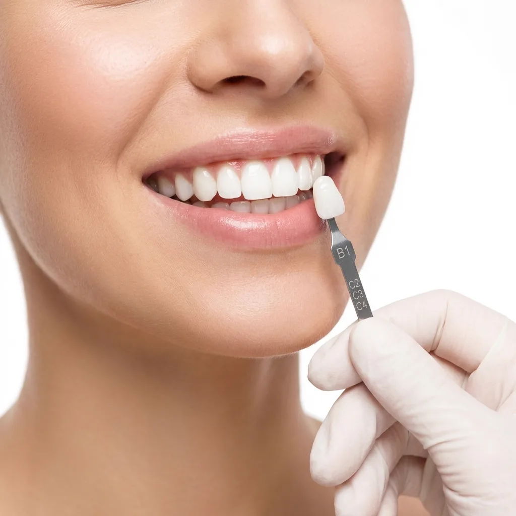
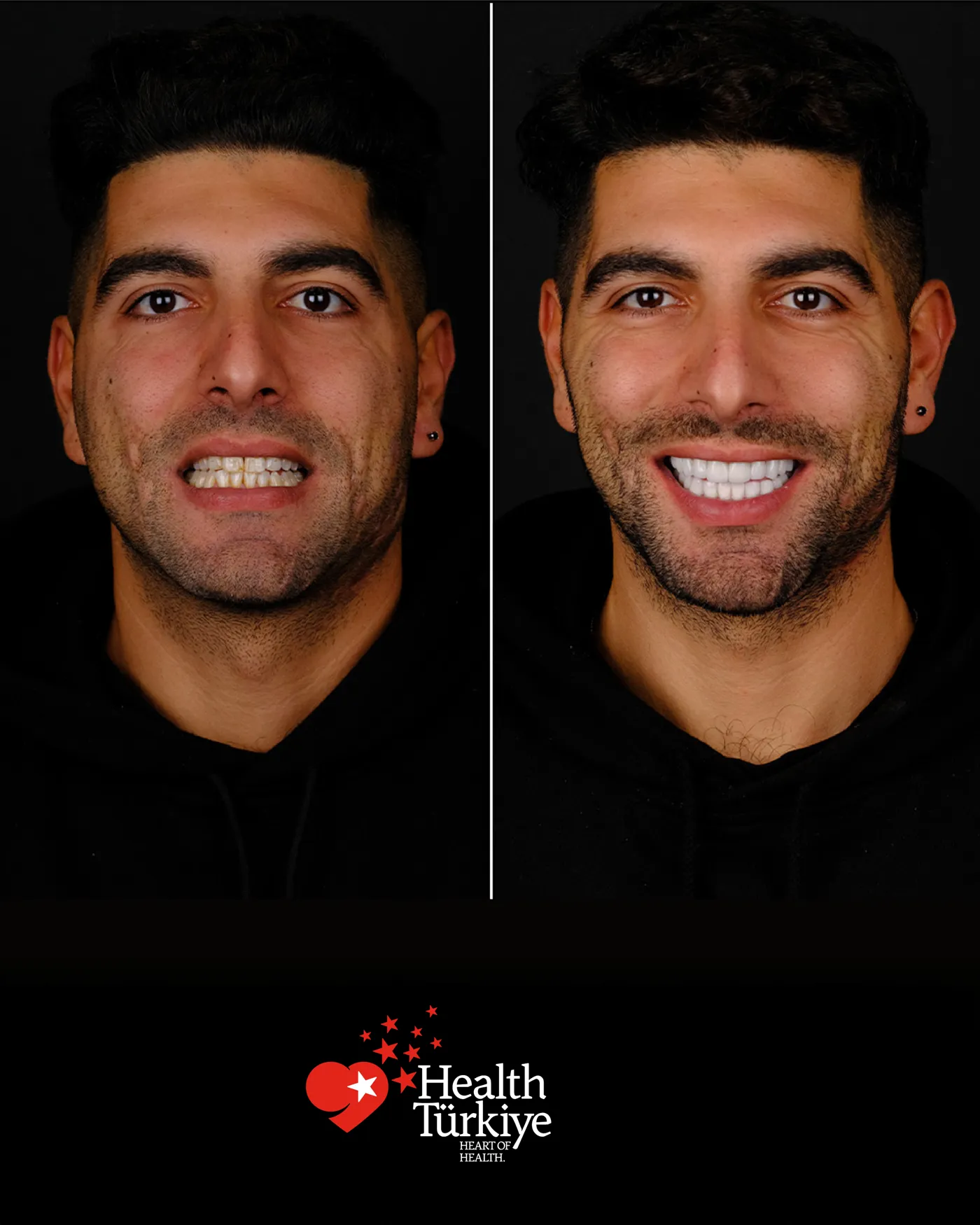
The same teeth look entirely different at different gum levels.
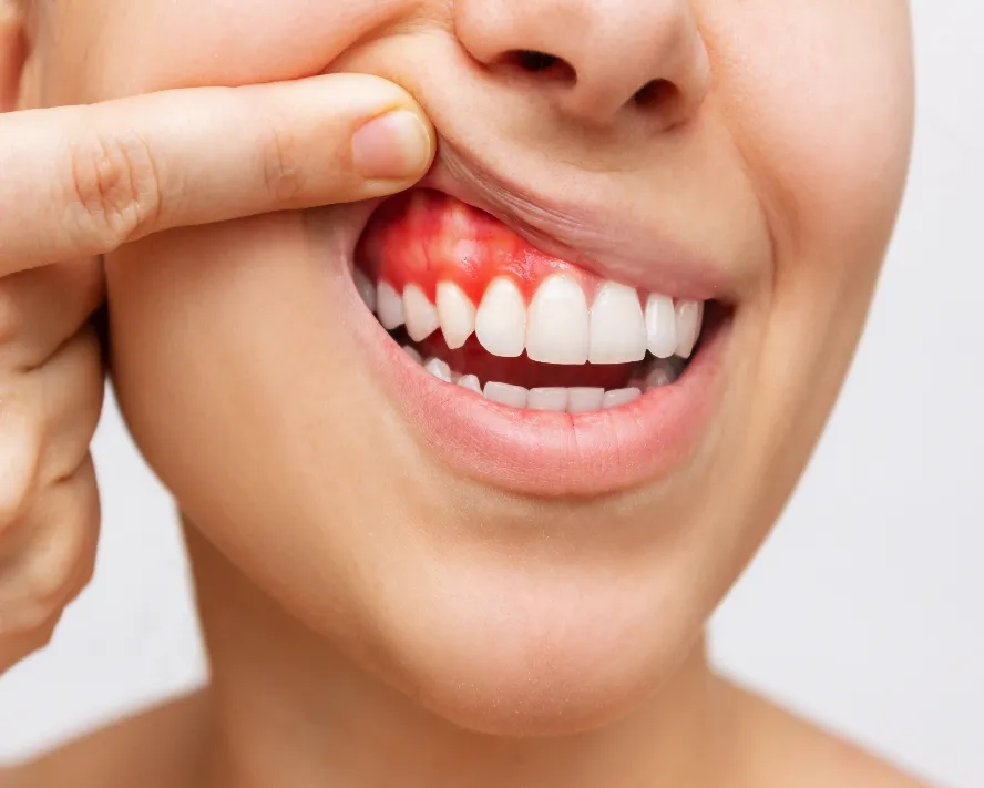
When too much gum shows
There is no single cause: excess gum over the teeth, an upper lip that lifts too far, or vertical overdevelopment of the upper jaw. The distinction matters because each needs a different approach.
An asymmetric gum line
If the gum levels of the front teeth differ, the smile looks unbalanced even when the teeth are equal in length. Crucially, crowning does not fix this — a crown changes the visible part of the tooth, not the gum line.
Black triangles between teeth
The triangular gap below the contact point is normally filled by the gum papilla. When it recedes the gap appears, usually because of underlying bone loss — so what looks like an aesthetic complaint is really a periodontal finding.
Getting Here, Booking and Emergencies from Liman
Route, booking and emergency arrangements for patients from Liman.
Getting here from Liman
Liman lies in the Konyaaltı district of Antalya; Konyaaltı is one of the central districts, about 8 kilometres from the clinic. We also see patients from neighbouring areas that share the route from Liman, such as Arapsuyu, Demircilik, Gürsu, Kuşkavağı. You can ask for the location on WhatsApp when travelling from Liman.
Appointments for Liman
At this distance, splitting treatment into visits is no problem for patients from Liman, and attending reviews is easy too. As with patients from Konyaalti generally, appointments for families from Liman can be scheduled consecutively.
Emergencies from Liman
Setting out from Liman at night is quick; come without delay in an emergency, and calling as you leave means preparation is done in advance. For patients coming from Dental Clinic in Liman, the clinic is open 24/7 and in an emergency the order is set by severity rather than arrival time. For spreading facial swelling, difficulty swallowing or breathing, or toothache with fever, go to the nearest emergency department.
Jaw Cysts: Growths That Enlarge Silently
Most cysts developing within the jawbone cause no pain and are found incidentally on a radiograph taken for another reason. The value of periodic radiographs therefore goes beyond looking for decay.
Where they arise
There are two main groups:
- Odontogenic: a cyst developing at the root tip of a non-vital tooth
- Developmental: a cyst surrounding the crown of an impacted tooth
- Impacted wisdom teeth are the most common source
- Rarely, those developing from embryonic tissue remnants
Why they should not be left
A cyst grows slowly but continuously, resorbing the bone around it. Once large enough it causes swelling of the jaw, can resorb the roots of neighbouring teeth, displace teeth, and thin the bone so much that a minor blow can fracture the jaw. What could be removed early with a small procedure requires extensive surgery when left.
Treatment and follow-up
Small cysts are removed completely and the cavity fills with bone on its own, with a graft placed if needed. Very large cysts may first be decompressed and allowed to shrink before removal. The tissue removed is always sent for pathological examination. Some types tend to recur, so annual radiographic follow-up is planned.